Live Cell Imaging of Mitochondria and Actin Dynamics During Synaptogenesis
In vitro and in vivo studies have shown that neurons can undergo neuritogenesis and synaptogenesis in response to various compounds that are being tested in the clinic for treating various neurological diseases. It is speculated that these synaptogenic activities lead to the formation of new neural circuits and corresponding therapeutic benefits. However, the mechanisms by which these compounds exert these effects (and their scope) remain poorly understood. Understanding the specific cellular changes that occur during synaptogenesis will provide important insights. I hypothesize that mitochondria play a central role in induced formation of new neurites and synapses, and that mitochondria and synapses are coupled via the actin cytoskeleton. To test these hypotheses, I will utilize unique imaging tools we developed to measure real-time mitochondria and actin dynamics in living neurons. I will use pharmacological and novel genetic tools to identify the role of the actin cytoskeleton in regulating mitochondria dynamics in live neurons. Multi-omics data will be collected to gain a complete understanding of global changes to cells during perturbations to the actin cytoskeleton. Together, these analyses will provide the most detailed assessment to date of the role of actin-mitochondria coupling during synaptogenesis, laying the foundation for improved understanding and treatment of a broad range of mental health disorders.
Grant Recipients

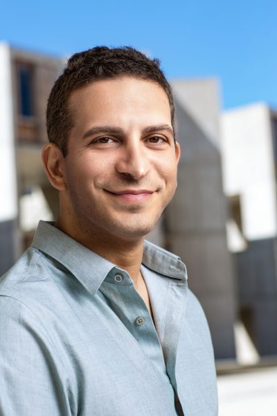
Dr. Uri Manor
Uri Manor is an Assistant Research Professor, Assistant Director of the Waitt Advanced Biophotonics Center, and Director of the Waitt Advanced Biophotonics Core. Uri develops and applies artificial intelligence-based computational approaches (deep learning) that integrate data from optical and electron microscopy techniques increasing image resolution, sensitivity, and collection speed beyond what’s possible with any individual method. Uri’s current research focuses on developing novel artificial intelligence approaches to increase the resolution, sensitivity and speed of the next generation of microscopes, as well as designing nanoprobes for high spatiotemporal resolution imaging of subcellular dynamics. His main biological interests are mitochondria, hearing loss, neurodegeneration and synaptic plasticity.
Prior to joining Salk, Uri did his PhD thesis research work with Bechara Kachar (NIH), and his postdoctoral training with Jennifer Lippincott-Schwartz (NIH and Janelia Farms) using advanced quantitative imaging approaches, such as super-resolution and live cell imaging, automated analysis and segmentation of microscopy data, and computational modeling of biophysical and biochemical dynamics in the cell.
